H&E Staining Protocol / Oil Red O And Hematoxylin And Eosin Staining For Quantification Of Atherosclerosis Burden In Mouse Aorta And Aortic Root Springerlink / Hematoxylin and eosin stain (h&e) is one of the principal stains in histology.
H&E Staining Protocol / Oil Red O And Hematoxylin And Eosin Staining For Quantification Of Atherosclerosis Burden In Mouse Aorta And Aortic Root Springerlink / Hematoxylin and eosin stain (h&e) is one of the principal stains in histology.. Hematoxylin and eosin stain (h&e) is one of the principal stains in histology. Do not allow them to dry or thaw prior to being put in the ethanol. Primary differences are dye composition, staining protocol, and intensity of blue dye. This is the standard reference stain used in the study of histochemical tissue pathology. Hematoxylin & eosin (h & e) stain protocol principle:
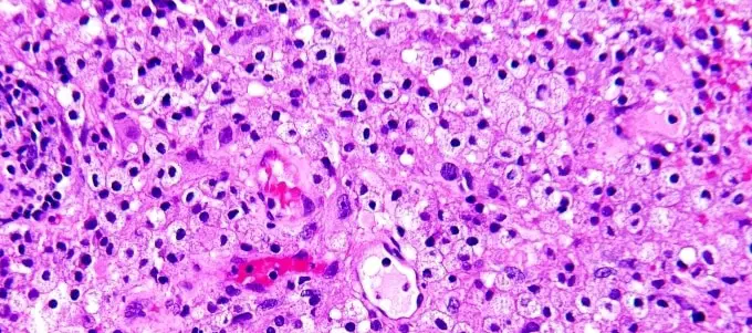
H&e stain nucleus in blue and cytoplasm in red, therefore allowing for morphological analyses, such as myofiber diameter. The timing of these steps will need to be optimized for your experiments, but the details in these protocols provide a guideline. H&e stain, he stain or hematoxylin and eosin stain, is a popular staining method in histology. In histopathology laboratory, the hematoxylin and eosin stain is referred to as the routine stain. These stain pink as a result.
Background Tumor Proliferation Assessment Challenge 2016 from tupac.tue-image.nl Hematoxylin is not even synthetically produced, but is instead extracted from the logwood tree. This protocol is applied in the routine staining of cationic and anionic tissue components in tissue sections. Progressive, modified progressive, and regressive. Cell staining is a technique used for the main purpose of increasing contrast through changing the color of some of the parts of the structure being observed thus allowing for a clearer view. H&e stain or he stain) is one of the principal tissue stains used in histology. Primary differences are dye composition, staining protocol, and intensity of blue dye. H & e stain (hematoxylin and eosin) harris hematoxylin* is not recommended. There are typically three types of h&e stains:
Primary differences are dye composition, staining protocol, and intensity of blue dye.
Especially during the summer months, be sure to not let the slides set out overnight/extended period of time. Distilled and tap water 5. Hematoxylin and eosin stain or haematoxylin and eosin stain (h&e stain or he stain) is one of the principal stains in histology. As its name suggests, h&e stain makes use of a combination of two dyes, namely hematoxylin and eosin. H&e stain nucleus in blue and cytoplasm in red, therefore allowing for morphological analyses, such as myofiber diameter. The next step to having a good stain is determining what type of h&e stain is desired. Hematoxylin and eosin stain or haematoxylin and eosin stain (often abbreviated as: | special stains and h & e 1. Any suggestions on h & e staining protocol for frozen sections using automatic stainer. I'm looking into h&e protocols for staining a cytospin slide of peripheral blood mononuclear cells. Hematoxylin & eosin (h & e) stain protocol principle: Protocol and troubleshooting tips for whole mount immunohistochemical staining. Introductionhematoxylin and eosin (h&e) stains have been used for at least a century and are still essential for recognizing various tissue types and the morphologic changes that form the basis of contemporary cancer diagnosis.
5 external links o 5.1 protocol. There are typically three types of h&e stains: Can anyone help me with. Protocol and troubleshooting tips for whole mount immunohistochemical staining. Gill's and mayer's formulations are suitable.
Hematoxylin And Eosin Staining Principle Procedure And Interpretation Laboratoryinfo Com from laboratoryinfo.com Hematoxylin and eosin stain or haematoxylin and eosin stain (h&e stain or he stain) is one of the principal stains in histology. There are a variety of stains that can be used in microscopy. | special stains and h & e 1. Especially during the summer months, be sure to not let the slides set out overnight/extended period of time. The staining will get more intense after time, be sure not to oversaturate section. H & e stain (hematoxylin and eosin) harris hematoxylin* is not recommended. Of the principal stains in histology. This protocol is applied in the routine staining of cationic and anionic tissue components in tissue sections.
Hematoxylin & eosin (h & e) stain protocol.
This protocol describes h&e staining of tissue and cell sections. 5 external links o 5.1 protocol. The staining will get more intense after time, be sure not to oversaturate section. This is the standard reference stain used in the study of histochemical tissue pathology. Follow the appropriate staining protocol described below. Hematoxylin is not even synthetically produced, but is instead extracted from the logwood tree. This protocol is applied in the routine staining of cationic and anionic tissue components in tissue sections. Gill's and mayer's formulations are suitable. Check out the principle, procedure, and results on home. Rosen lab, department of molecular and cellular biology, baylor college of medicine) step by step protocol. A first person view of how to manually stain slides using the h&e staining method.the protocol demonstrates use of ehrlich's hematoxylin recipe which is. Hematoxylin and eosin stain (h&e) is one of the principal stains in histology. Any suggestions on h & e staining protocol for frozen sections using automatic stainer.
The staining will get more intense after time, be sure not to oversaturate section. | special stains and h & e 1. H & e stain (hematoxylin and eosin) harris hematoxylin* is not recommended. Hematoxylin & eosin (h & e) stain protocol principle: 3 x 3´ xylene (blot excess xylene before going into ethanol) 3 x 3´ 100% ethanol 1 x 3´ 95% ethanol 1 x 3´ 80% ethanol 1 x 5´ deionized h2o •while sections are in.
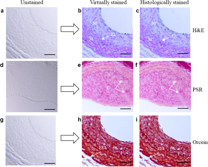
Deep Learning For Virtual Histological Staining Of Bright Field Microscopic Images Of Unlabeled Carotid Artery Tissue Springerlink from media.springernature.com For example when a pathologist looks at a biopsy of a suspected cancer. The staining will get more intense after time, be sure not to oversaturate section. Progressive staining occurs when the hematoxylin is added to the tissue without being. In histopathology laboratory, the hematoxylin and eosin stain is referred to as the routine stain. The next step to having a good stain is determining what type of h&e stain is desired. This combination deferentially stains various tissue the principle behind h & e stain is the chemical attraction between tissue and dye. I'm looking into h&e protocols for staining a cytospin slide of peripheral blood mononuclear cells. H & e stain (hematoxylin and eosin) harris hematoxylin* is not recommended.
H&e stain, he stain or hematoxylin and eosin stain, is a popular staining method in histology.
Hematoxylin & eosin (h&e) staining protocol. Hematoxylin and eosin stain or haematoxylin and eosin stain (h&e stain or he stain) is one. Primary differences are dye composition, staining protocol, and intensity of blue dye. Here we describe a protocol to examine muscle histology and myofiber types using hematoxylin and eosin (h&e) and immunofluorescence staining, respectively. H&e stain, he stain or hematoxylin and eosin stain, is a popular staining method in histology. Protocol and troubleshooting tips for whole mount immunohistochemical staining. Progressive, modified progressive, and regressive. 5 external links o 5.1 protocol. Hematoxylin & eosin (h & e) stain protocol. Hematoxylin is not even synthetically produced, but is instead extracted from the logwood tree. There are typically three types of h&e stains: Having difficulty with uniform staining. Representative h&e and ihc stains in pancreatic sections.
You have just read the article entitled H&E Staining Protocol / Oil Red O And Hematoxylin And Eosin Staining For Quantification Of Atherosclerosis Burden In Mouse Aorta And Aortic Root Springerlink / Hematoxylin and eosin stain (h&e) is one of the principal stains in histology.. You can also bookmark this page with the URL : https://kanznoy.blogspot.com/2021/05/h-staining-protocol-oil-red-o-and.html
Share Awesome
Belum ada Komentar untuk "H&E Staining Protocol / Oil Red O And Hematoxylin And Eosin Staining For Quantification Of Atherosclerosis Burden In Mouse Aorta And Aortic Root Springerlink / Hematoxylin and eosin stain (h&e) is one of the principal stains in histology."

Belum ada Komentar untuk "H&E Staining Protocol / Oil Red O And Hematoxylin And Eosin Staining For Quantification Of Atherosclerosis Burden In Mouse Aorta And Aortic Root Springerlink / Hematoxylin and eosin stain (h&e) is one of the principal stains in histology."
Posting Komentar